
The Mystery of the 20th Century Epidemic
In the late 1700s and 1800s, the medical literature of the Western world was remarkably silent regarding the devastating cognitive decline we now call Alzheimer’s Disease (AD). The pathologists of the Victorian era were not mere hobbyists; they were meticulous observers who cataloged rare conditions with obsessive detail. It is a profound historical anomaly that they would have overlooked a condition characterized by the obvious morphological markers of AD—shrunken brain volume, distinct plaques, and the total dissolution of the patient’s persona. Yet, the first clinical case of AD was not identified until 1903, appearing on the heels of the 19th-century dental revolution that established “silver” mercury amalgams as a standard medical material.
Today, that silence has been replaced by the roar of a global epidemic. Alzheimer’s has become our most feared neurological specter, a disease of “unknown etiology” that remains a riddle despite billions of dollars in research. We walk through modern memory care units and see the human cost—the “long goodbye” that strips families of their history. We are told this is a natural byproduct of an aging population, a genetic inevitability of living longer. But the chronological correlation between the rise of mercury use and the explosion of AD suggests a more sinister origin.
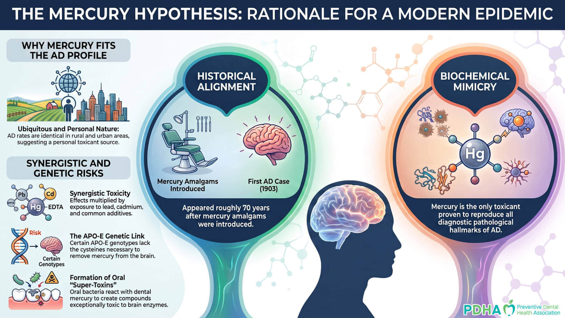
Dr. Boyd E. Haley, a professor of chemistry and specialist in biochemical research, has spent decades studying the biochemical effects of mercury and other toxicants. In his 2007 paper, The Relationship of the Toxic Effects of Mercury to Exacerbation of the Medical Condition Classified as Alzheimer’s Disease, Haley presents a provocative hypothesis: that Alzheimer’s disease may not simply be a natural consequence of aging, but may be driven or exacerbated by chronic mercury exposure. Rather than viewing the hallmark changes associated with Alzheimer’s solely as causes of dementia, Haley challenges readers to consider whether they may instead be biochemical evidence of an underlying toxic process.
“...for any toxicant, or class of toxicants, to be proposed as involved in the etiology of AD it must be available almost equally to individuals living in markedly different locations. The toxicant proposed must also explain the genetic susceptibility aspects of AD. Further, under experimental conditions the putative toxicants must be able to cause or exacerbate many of the biochemical abnormalities found in the AD brain.”
Dr. Boyd E. Haley
How Mercury Replicates Every Diagnostic "Hallmark" of Alzheimer’s
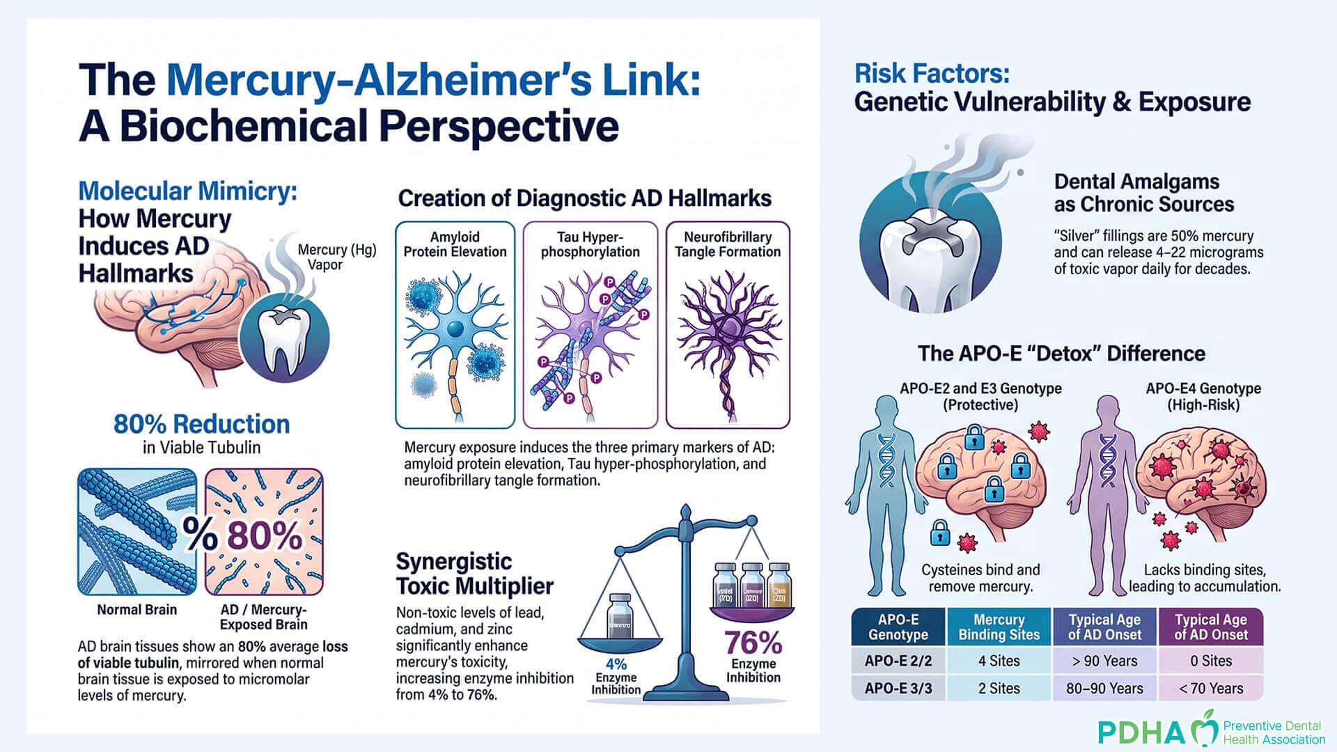
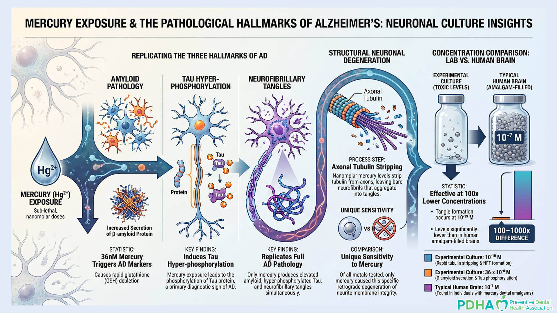
In the diagnostic theater of neurology, Alzheimer’s is identified by three pathological footprints: elevated amyloid protein, the hyper-phosphorylation of Tau, and the formation of neurofibrillary tangles (NFTs). For decades, research has focused on clearing these markers as if they were the primary invaders. However, Dr. Haley’s research reveals that mercury (Hg2+) is the only toxicant known to science that can reproduce all three of these diagnostic markers in appropriate test systems. Mercury doesn’t just damage the brain; it mimics the disease so perfectly that the two become indistinguishable at the cellular level.
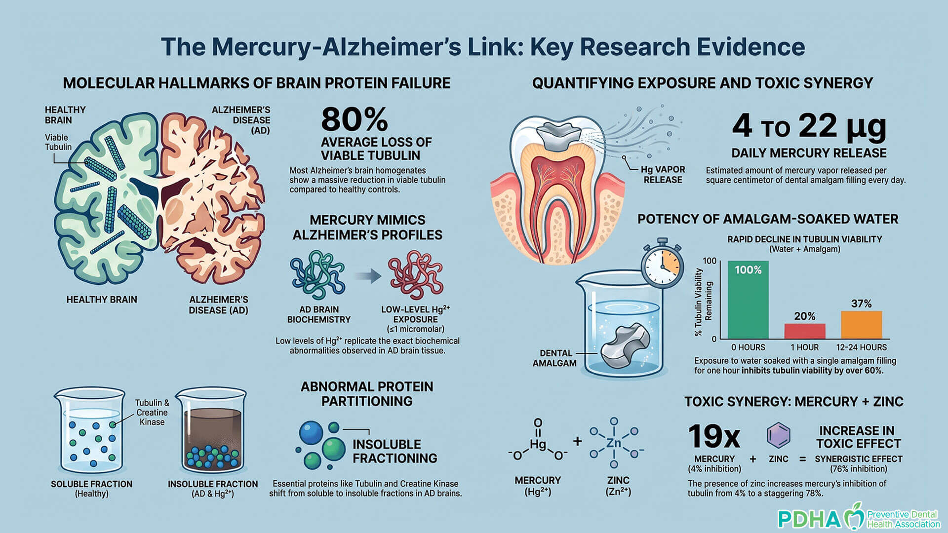
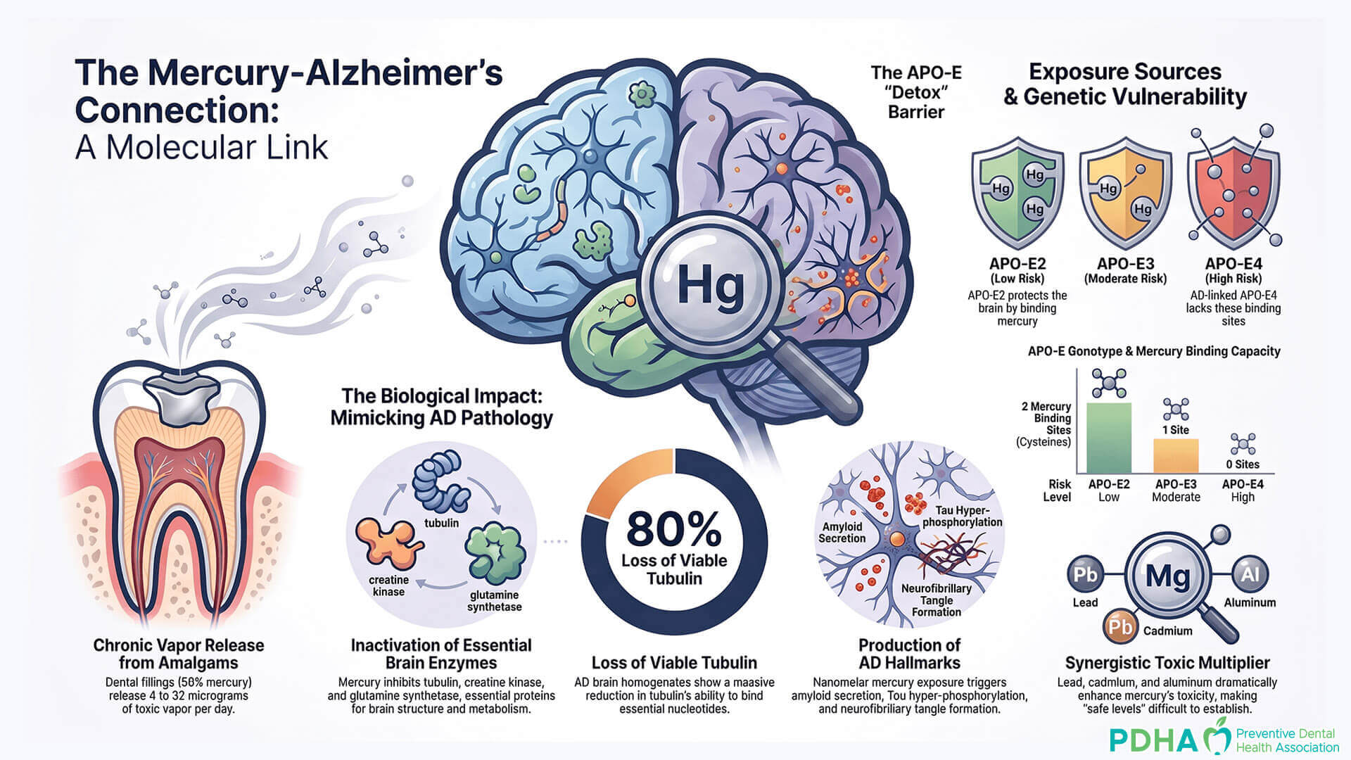
The mechanism of this destruction is a masterclass in biochemical efficiency. Mercury targets thiols (sulfhydryls), which are the essential “anchors” for brain enzymes. Think of these enzymes as the essential infrastructure of a city. Creatine kinase (CK) is the power grid, responsible for energy metabolism; glutamine synthetase (GS) is the waste management system, regulating toxic neurotransmitters like glutamate. Mercury inactivates both, plunging the neuron into a state of energetic failure and toxic buildup. But its most visual crime occurs within the neuron’s structural scaffolding: tubulin.
In a landmark study involving snail neurons, researchers demonstrated that when neurons are exposed to even nanomolar levels of mercury—levels far lower than those found in the average human brain—a “rapid stripping” occurs. The tubulin, which maintains the structural integrity of the axons, is essentially dissolved. In a striking visual collapse, the axons wither, leaving behind bare, aggregated neurofibrils. These collapsed structures are the very neurofibrillary tangles that define the Alzheimer’s brain. As Dr. Haley summarizes:
"The rapid inactivation of the brain thiol-sensitive enzymes (tubulin, creatine kinase and glutamine synthetase) occurs after: (a) the addition of low micromolar levels of Hg2+, (b) exposure to mercury vapor (Hg0) or (c) the addition of Thimerosal."
Dr. Boyd E. Haley
The Constant Toxic Vapor Inches from the Brain
The primary source of this chronic exposure is a scientific misnomer that sits inches from the human brain. Dental amalgams, marketed as “silver fillings,” are actually composed of approximately 50% elemental mercury. A single large filling may contain an entire gram of mercury—one million micrograms. To appreciate the scale of this “retention toxicity,” consider that if a filling lost a toxic dose of 10 micrograms per day, it would take nearly 300 years to exhaust its supply. Most patients carry these “toxic reservoirs” for their entire adult lives.
The regulatory shield protecting amalgams relies on a structural flaw in testing: the use of blood and urine levels to determine safety. Mercury is not a transient traveler in the bloodstream; it is a “retention toxicant.” Approximately 80% of inhaled mercury vapor is absorbed and stored in fatty tissues like the brain and heart. Using a blood test to measure mercury burden is like trying to judge the amount of trash in a landfill by looking at the exhaust of the garbage truck—it misses the massive accumulation in the tissues. The most chilling evidence of this retention comes from studies of idiopathic dilated cardiomyopathy (IDCM), where victims were found to have mercury levels in their heart tissue 22,000 times higher than controls who died of other heart diseases.
There is a deep irony in the “safety” data provided by manufacturers. Dr. Haley highlights a study by Chew et al. that intended to show the stability of amalgams using a brand called Composil, which was marketed specifically as a “non-mercury-releasing” material. The results were a self-indictment of the industry. Even this “safe” version was found to be leaking significant amounts of toxin. As the study noted:
“The overall mean release of mercury was 43.5 +/- 3.2 micrograms/cm2/24 hr, and the amount of mercury released remained fairly constant during the duration of the experiment (2 years).”
APO-E4 as a Detoxification Failure
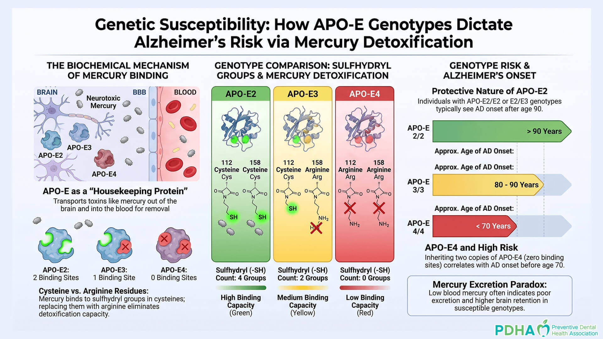
Modern medicine has largely accepted the APO-E4 genotype as a genetic “death sentence.” Those with two copies of the gene are warned of a high probability of early-onset Alzheimer’s. However, the mercury hypothesis transforms APO-E4 from a “disease-causing gene” into a “detoxification failure.” In the central nervous system, the APO-E protein acts as a specialized filter or “housekeeping” agent, designed to bind to toxins and carry them across the blood-brain barrier for disposal.
The functional difference between the “protective” APO-E2 and the “risky” APO-E4 is purely chemical. APO-E2 contains two mercury-binding cysteines (sulfhydryl-containing amino acids) that act like chemical magnets to haul mercury out of the brain. APO-E3 has only one of these sites. APO-E4, tragically, has zero. This is not a gene that “causes” plaques; it is a gene that fails to remove the toxicant that induces those plaques.
This shift in perspective is ethically transformative. If an individual has a “poor filter” gene, the danger is only realized if the environment is “dirty.” In a world without mercury exposure from amalgams or vaccines, the lack of mercury-binding sites on the APO-E4 protein would be biologically irrelevant. By labeling APO-E4 a “cause” of Alzheimer’s, we effectively blame the victim’s genetics for an environmental failure we refuse to address.
The Synergistic Death Trap: Why "Safe Levels" are a Scientific Myth
Regulatory agencies typically determine “safe” exposure levels by testing a single chemical on healthy rats in a controlled lab. This is a scientific fantasy. In the real world, humans do not live in isolation; we live in a chemical soup. Dr. Haley’s research demonstrates that mercury’s toxicity is not merely additive – it is synergistic. When mercury is combined with other metals or common substances, its lethality doesn’t just increase; it skyrockets.
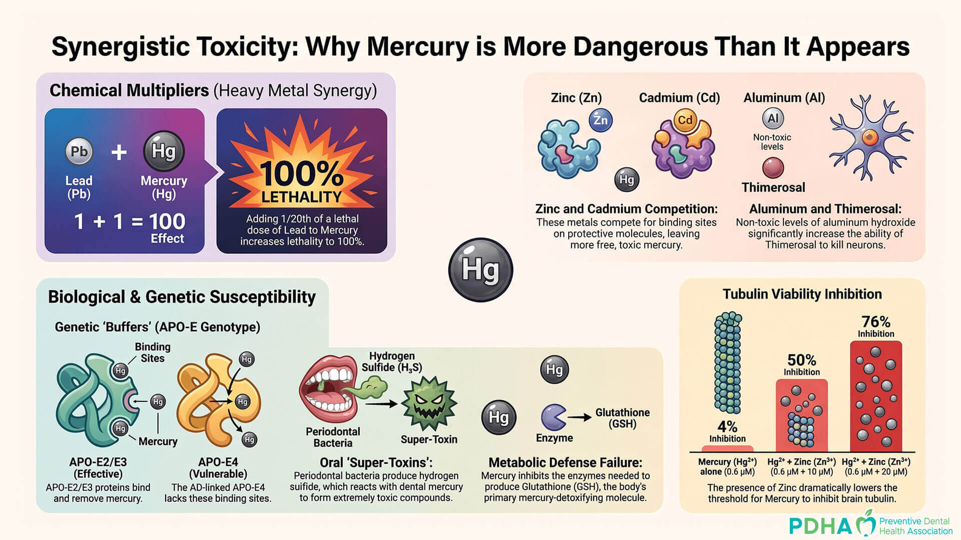
The data on this synergy is nothing short of terrifying. In controlled animal studies, researchers found that combining 1/20th of a lethal dose of lead (Pb2+) with a dose of mercury so low it caused zero deaths resulted in 100% lethality in the test group. This “synergistic death trap” is further compounded by daily life. The source context notes that common food additives like EDTA, antibiotics like tetracycline, and even the consumption of milk can dramatically increase mercury retention. Furthermore, the aluminum hydroxide found in vaccines has been shown to exponentially increase the neurotoxicity of mercury compounds like Thimerosal.
This reality renders the search for a “safe level” of mercury fundamentally flawed. A dose of mercury that might be “safe” for a laboratory rat eating monitored food is potentially devastating for a human who smokes (exposure to cadmium), has leaded-gasoline fumes in their history, and takes a course of antibiotics. When we calculate safety based on isolated variables, we ignore the synergy of modern life.
The "Super-Toxins" of the Oral Cavity
The danger of dental mercury is not limited to the vapor itself; it involves a secondary chemical reaction with the bacteria already present in our mouths. When a patient suffers from periodontal disease, anaerobic bacteria produce metabolic gases like hydrogen sulfide and methyl thiol. When these gases meet the mercury vapor from a “silver” filling, they create “super-toxins” known as methylthiomercury and dimethyl-thio-mercury.
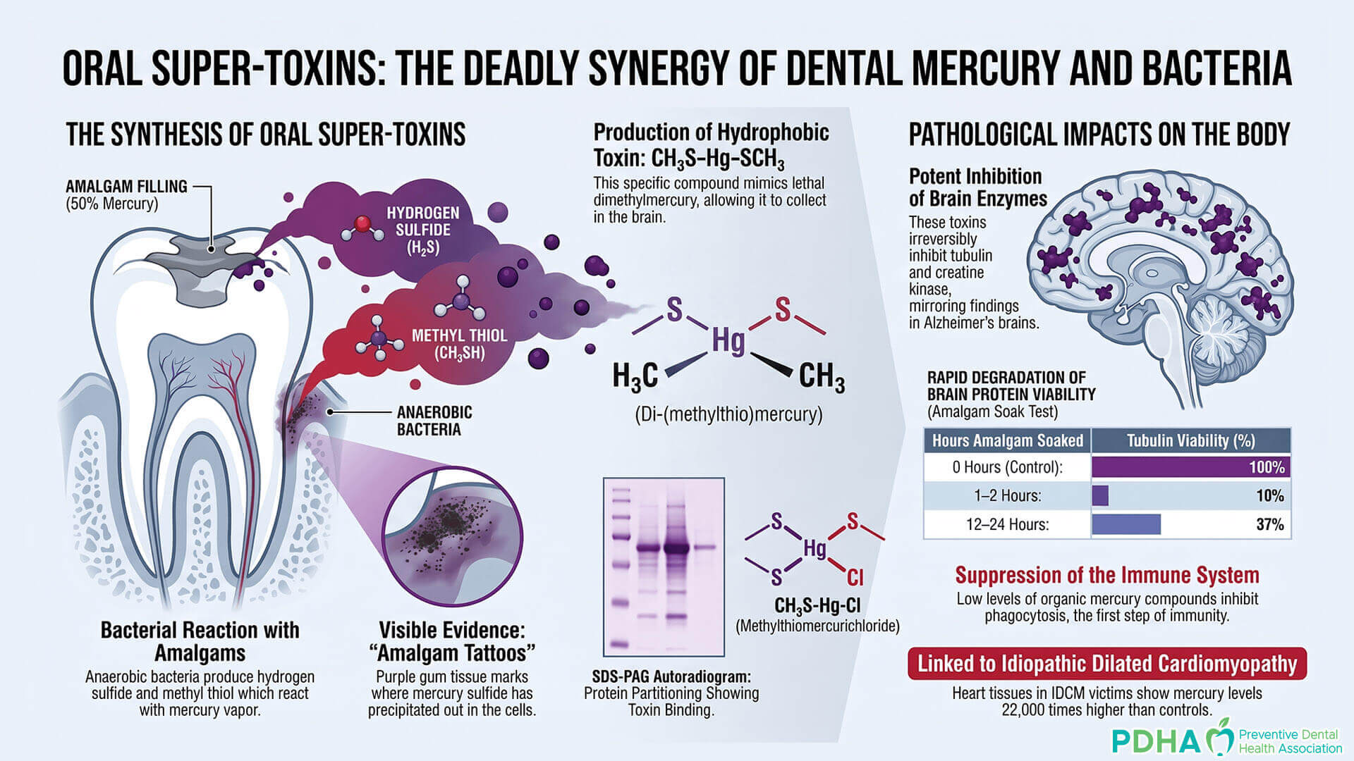
These compounds are exceptionally hydrophobic (fat-soluble), allowing them to slip through cellular membranes and the blood-brain barrier with ease. Their chemical profile is hauntingly similar to dimethylmercury—the compound that famously killed a Dartmouth chemistry professor after just a few drops penetrated her latex gloves. The presence of these toxins is often visible in the form of “amalgam tattoos,” the dark discoloration of the gums where mercury has reacted with bacterial sulfides.
If these “super-toxins” are precipitating in the gums, they are almost certainly circulating systemically. This brings us back to the “smoking gun” of heart tissue research. The massive mercury accumulation found in the hearts of IDCM patients suggests that these oral reactions are creating compounds that the body simply cannot excrete. We have allowed the oral cavity to become a chemical laboratory, synthesizing compounds of extreme lethality inches away from our central nervous system.
A Forward-Looking Reflection
The biochemical evidence presented by Dr. Boyd Haley suggests that we have been looking at the Alzheimer’s epidemic through the wrong end of the telescope. The amyloid plaques and neurofibrillary tangles that have been the targets of our “cure” efforts for decades are likely not the cause of the disease, but the scorched earth left behind by mercury-induced damage. From the structural stripping of neurons to the failure of the APO-E4 detoxification system, mercury is the only factor that explains every facet of the disease.
This leaves us with a profound ethical question: If we have the biochemical proof that mercury mimics every hallmark of our most feared neurological disease, why is the “silver filling” still the standard of care? We can no longer afford to view Alzheimer’s as an unavoidable mystery of the aging process. It is time to confront the possibility that we have traded our long-term neurological health for a cheap, century-old dental material, and in doing so, we have created an epidemic of our own making.
Watch: Mercury Causes the Biochemical Hallmarks of Alzheimer’s Disease
Boyd Haley, PhD
About the Preventive Dental Health Association
The PDHA is a non-profit organization dedicated to advancing biocompatible, science-based preventive oral health strategies that support systemic health and longevity. Additional information is available at thepdha.org.