Dental cavities are more than just a childhood rite of passage; they are a global health crisis. According to the Global Burden of Disease study, dental caries rank as the most widespread condition affecting permanent teeth, with approximately 560 million children impacted worldwide. For decades, the standard public health response has been the “silver” filling. But as we move further into the 21st century, a pressing question emerges: what actually happens to the mercury in those fillings once they are packed into a child’s mouth?
A landmark 2025 study by Macías-Lamas et al., conducted at the Universidad de Guadalajara in Mexico, has pulled back the curtain on the “oxidative genomic damage” occurring in pediatric patients. The findings are a wake-up call for the dental community, suggesting that the mercury released from these restorations isn’t just sitting there—it’s actively rewriting the cellular health of the next generation.
A landmark 2025 study by Macías-Lamas et al., entitled “Oxidative genomic damage in pediatric patients exposed to mercury released by dental amalgam”
The "50% Weight" Reality
In public health journalism, we often talk about “hidden in plain sight.” The term “silver filling” is a perfect example. These restorations are actually dental amalgam, and mercury isn’t just a trace ingredient—it constitutes 50% of the material’s total weight. This creates a startling tension in medical policy: one of the World Health Organization’s top ten pollutants of concern is being placed directly into the mouths of children as a primary restorative tool.
This reliance persists because amalgam is inexpensive and somewhat durable (if even at the expense of long term tooth health and structure). However, the study reminds us that mercury is lipophilic and volatile; it is constantly released through daily abrasion and corrosion, entering the bloodstream and oral tissues.
“The World Health Organization (WHO) includes mercury (Hg) among the ten pollutants of particular concern for public health… Hg constitutes the primary component of this type of amalgam, making up 50% of its weight.”
Visible Damage in Just 15 Days
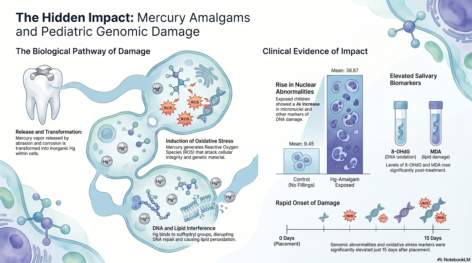
The most alarming journalistic “lead” from this research is the sheer speed of biological change. Using the Buccal Micronucleus Cytome (BMcyt) assay—a method of analyzing cells scraped from the inner cheek—researchers detected a significant rise in “nuclear abnormalities” (NAs) just 15 to 30 days after fillings were placed.
These aren’t just minor cellular glitches. These abnormalities, including micronuclei and nuclear buds, are markers of chromosomal instability. The study makes the gravity of this clear: an increase in these biomarkers is directly associated with genotoxic processes and a higher risk of developing cancer later in life. This isn’t a long-term “wait and see” scenario; it is short-term exposure leading to immediate, visible markers of long-term risk.
Saliva as a Mirror for Internal Stress
The researchers used saliva as a biological mirror to reflect the “invisible” stress occurring within the children. They tracked two specific biomarkers: 8-hydroxy-2′-deoxyguanosine (8-OHdG), a tell-tale sign of DNA oxidation, and Malondialdehyde (MDA), which indicates lipid peroxidation—essentially the “rusting” of cell membranes.
As mercury vapor is absorbed by the oral mucosa, these markers spike. This correlation proves that the mercury isn’t just a localized resident of the tooth; it is a systemic driver of oxidative stress that can attack genetic material and cell integrity simultaneously.
“Individuals exposed to Hg-containing dental amalgam exhibit increased NAs in oral epithelial cells, as well as increased levels of 8-OHdG and MDA in saliva, which are directly related to genotoxicity, oxidative DNA damage and lipid peroxidation.”
The Body’s Broken Repair Kit
To understand why mercury is so insidious, we have to look at the “Base Excision Repair” (BER) systems—the body’s natural mechanic for fixing broken DNA. Mercury utilizes a form of “molecular mimicry” to sabotage these systems.
Once elemental mercury enters the body, it is transformed into an inorganic form by the enzyme hydrogen peroxide catalase. In this state, it has a ravenous affinity for “sulfhydryl groups.” It doesn’t just cause damage; it physically displaces zinc in the “zinc finger domains” of repair proteins. By kicking out the zinc, the mercury essentially breaks the repair kit. For a growing child with rapidly dividing cells, this “double hit”—increasing DNA damage while simultaneously blocking the pathways meant to fix it—is a profound biological gamble.
The "Pro-Oxidative" Baseline of Cavities
One of the study’s most nuanced findings involves the state of the child before the dentist even picks up a drill. Children with active cavities already show higher levels of oxidative stress and nuclear abnormalities. This is due to “microbial dysbiosis”—a bacterial imbalance—and the chronic inflammation that comes with tooth decay.
This means that many pediatric patients are already in a “pro-oxidative” state. Adding a mercury-containing amalgam to this environment acts as a chemical amplifier. It layers a known genotoxin on top of an already stressed system, significantly increasing the total burden of genomic stress compared to children with healthy teeth.
Conclusion: The Move Toward Genomic Safety
The Macías-Lamas study forces us to look beyond the surface of the tooth and toward the systemic horizon. We now know that mercury exposure is linked to a litany of “hidden” impacts: neurological and neurodegenerative disorders, renal (kidney) dysfunction, and cardiovascular diseases.
While dental amalgam has been a cost-effective workhorse for decades, we must weigh those savings against the biological cost of chromosomal instability and impaired DNA repair in children. With the rise of safer alternatives like composite resins and glass ionomers—materials that don’t carry this same genotoxic profile—we arrive at a crossroads.
Is it time for dental professionals and parents to move past the age of mercury and prioritize the long-term genomic health of the next generation? The cellular evidence suggests the answer is already in our mouths.